Karyogram Creation
You may benefit most from the following customizations:
- Accommodate a diverse array of banding techniques currently in use.
-
Utilize various specimens for banding analysis, including amniotic fluid, peripheral blood, chorionic villus, bone marrow, and tissue, without limitations based on specific diseases.
-
Incorporate numerous features to support your interpretation of metaphases and streamline your karyogram creation process.
-
Maintain a continuous log of processing steps and enjoy unrestricted access to the original images.
-
Facilitate smooth transitions between different capture settings, allowing for effortless shifts from brightfield to fluorescence and vice versa.
-
Choose the option for manual image acquisition with one-click capture, automatic contrast enhancement, and the selection of the optimal focus plane.
-
Creation of karyograms utilizing Deep Neural Networks (DNN) supersedes the machine learning tools used in earlier versions.
-
Chromosome separation in unprocessed metaphase images facilitated by DNN technology, with the goal of minimizing the need for manual intervention.
-
DNN-based chromosome assignment resulting in a karyogram draft that serves as a working basis for the adjustments and the evaluation performed by the experienced professional.
-
A more efficient workflow that has the potential to significantly decrease the amount of manual effort and time required to prepare the karyogram.
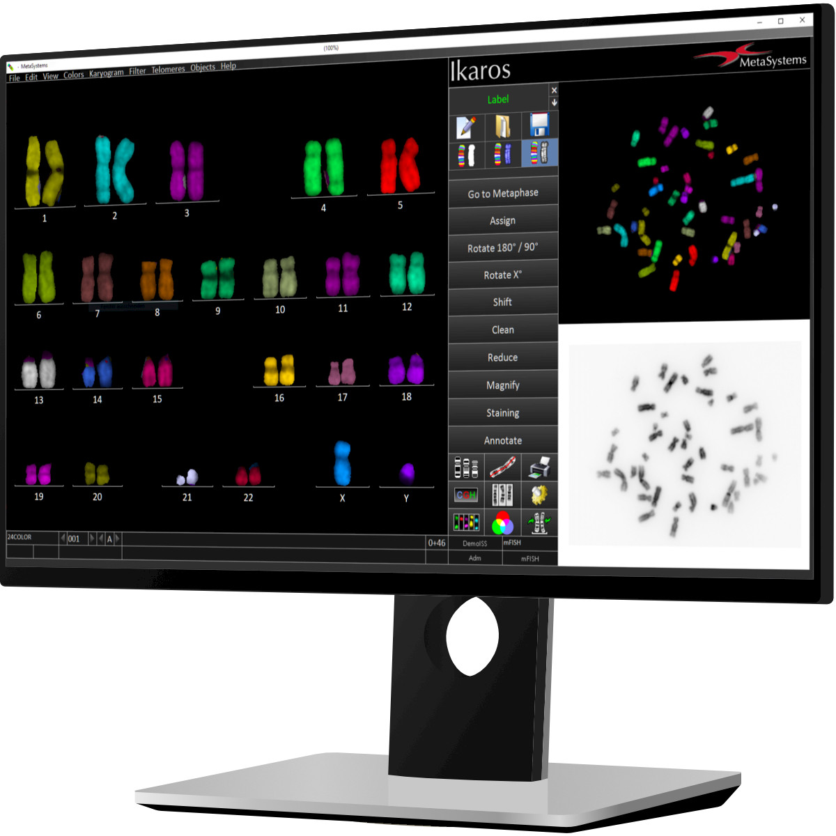
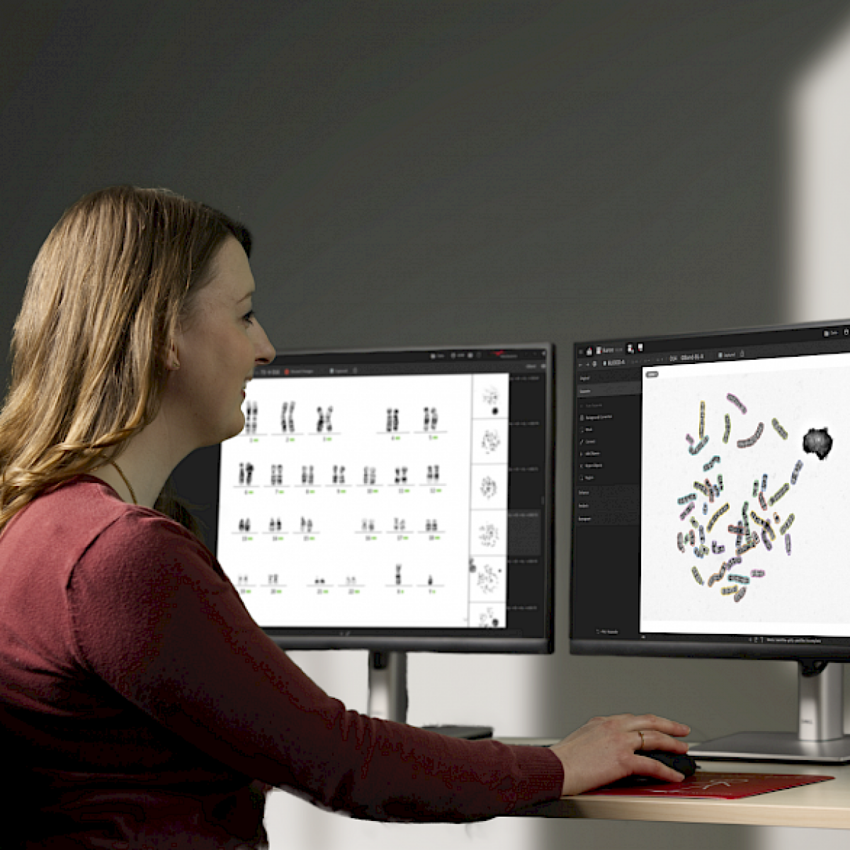
The Ikaros software integrates an intuitive graphical user interface with a range of potent processing tools, delivering the necessary flexibility in the process of karyogram creation. Engineered to reduce the number of interactions, Ikaros holds the potential to decrease the time needed for both analysis and result review compared to a manual workflow.
Ikaros offers tools to assist cytogeneticists in evaluating metaphases prepared using various prevalent chromosome banding methods (such as G-Banding and Q-Banding). It also supports diverse specimen types, including peripheral blood, bone marrow, amniotic fluid, and chorionic villi.
The latest versions of Ikaros by MetaSystems have integrated features based on Deep Neural Networks (DNNs), designed to assist cytogeneticists in segmenting metaphase chromosomes and classifying them within the karyogram. This cutting-edge technology supersedes the respective algorithms used in earlier versions of Ikaros, offering the potential to markedly decrease the number of interactions needed in comparison to the former workflow.
Like its predecessors, the latest versions of Ikaros support users in separating chromosomes in the metaphase. However, the adoption of a DNN-based workflow marks a notable progression in this process, aiming to lower the rate of segmentation errors and consequently lessen the need for manual corrections by the users.
This introduces the deployment of the new DNN-based tools as an additional means to achieve time savings in metaphase processing.
In the process of karyogram creation, another traditionally laborious task is the classification of chromosomes into their appropriate classes on the karyogram form. DNN technology also aids in this step, reducing the need for manual input and corrections. As a result, cytogeneticists are presented with an initial karyogram draft. This draft serves as a working basis for the adjustments and evaluation performed by the experienced professional.
FAQ
No. In the new Ikaros, the DNN capabilities facilitate the segregation of chromosomes and their categorization into the appropriate karyogram classes. This process yields a preliminary suggestion that requires further assessment and potential modification by a skilled user. Ultimately, the cytogeneticist is responsible for creating the final karyogram and conducting its analysis.
Yes, once Ikaros generates a karyogram proposal via its DNN features, users have the possibility and the responsibility to review and, if needed, amend this proposal. They have access to the same respective tools that have been present in earlier versions of Ikaros. Nevertheless, when an appropriate DNN is utilized, it is anticipated that the need for manual adjustments will be considerably reduced compared to the machine learning algorithms used in prior versions.
The response to this inquiry is contingent on various elements. Factors such as the nature of preparations, working methods employed, and the time allocated for result verification and reporting all play crucial roles in determining processing time. Nonetheless, insights from laboratories that have adopted the new feature indicate a noticeable decrease in case processing times, attributed to the diminished requirement for manual intervention in contrast to the algorithms previously in use.
No, there is no need for a replacement. Starting with version 6.3, every new version of Ikaros comes ready for DNN-based chromosome separation and classification and can be upgraded to include this feature. For efficient handling of DNN operations, an additional graphics card is needed to facilitate the computations. This card can be installed directly in the workstation or in a distinct system. For further information on the technical options available, please feel free to get in touch with us.
Chromosome analysis with the help of karyograms is central to cytogenetics, and Ikaros enhances this process, allowing the users to set up a versatile digital workflow. The software supports various banding techniques, specimen types, and advanced image processing tools, all within an intuitive, customizable interface.
The latest versions of Ikaros offer the option of implementing Deep Neural Networks (DNN) in user workflows, which help reduce segmentation errors, minimize manual corrections, and save time in metaphase processing. The DNN technology optimizes chromosome segmentation and classification and provides an initial karyogram draft as a basis for expert review. Delivering these innovative features while maintaining backward compatibility, Ikaros ensures that long-time users can seamlessly transition to the latest version and decide how much of this functionality they want to implement in their workflows.
Fluorescence Imaging
You may benefit most from the following customizations:
-
Effortlessly capture images with a single click, utilizing up to 12 color channels.
-
Get correct exposures at the first shot, thanks to automatic integration time.
-
Eliminate the need for time-consuming processing in a dark room.
-
Utilize a monochrome camera for the independent acquisition of single-color channels, automatically merging them to create a color image.
-
Refine and edit specific color channels or even individual areas within the image with selective editing capabilities.
-
View individual color separations and explore various pseudo-color visualizations.
-
Maintain a continuous log of processing steps and enjoy unrestricted access to the original images.
-
Opt for optional upgrades to enable support for color karyotyping, Multicolor FISH (mFISH) and the exclusive Multicolor Chromosome Banding (mBAND) method.
Moreover, Ikaros introduces a color fluorescence mode, displacing traditional photography and eliminating the time-consuming and laborious processing steps typically performed in a darkroom. Color images are produced by sequentially capturing monochrome images of the distinct color components and then automatically combining them to form a cohesive color image. The automated integration time control guarantees accurate exposures from the initial shot, eliminating the need for cumbersome test exposures or guesswork in determining the correct integration time. This not only reduces hands-on time but also extends the lifespan of the specimen. Even faint fluorescent signals against a strong counterstain result in clear and sharp images.
Legal Note
Ikaros 6.3 (SaMD + System)
Ikaros 6.3 is classified as an in vitro diagnostic (IVD) medical device in the European Union in accordance with the In Vitro Diagnostic Medical Device Directive 98/79/EC. It carries the CE label unless otherwise indicated and must be used within the scope of its intended purpose.
Ikaros is utilized in numerous countries worldwide. However, its use for clinical diagnostics depends on the regulatory requirements of each respective country or region.
Some hardware components supplied by third-party manufacturers are not included in the Ikaros IVD product and are therefore not part of the IVD medical device.